


Early detection, diagnosis and treatment are the keys to preventing vision loss from Macular Degeneration. It is often possible to detect early signs of Macular Degeneration during your regular eye examination, so scheduling regular eye exams is an excellent way to make an early diagnosis of Macular Degeneration. If you are over the age of 40-45 and you have a family history of Macular Degeneration, we recommend that you have a thorough eye examination, including a dilated retinal evaluation, each year. Please be sure to tell our staff if you have a family history of Macular Degeneration. It is also important to know the symptoms of Macular Degeneration. If at any time you experience “distortion” or “twisting”, “shadowing” or “bending” of objects in your vision, you should schedule an appointment immediately. If Macular Degeneration is diagnosed early enough, we are very fortunate to have a number of possible treatment options that may help to slow or even halt the progression of vision loss. However, patients must understand that once the macula has been damaged, there is no treatment that currently can reverse that damage and the associated loss of vision.
Dilated Retinal Exam
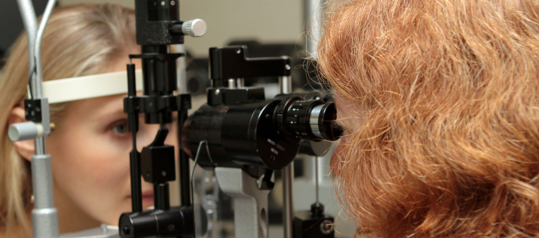
During your eye examination, eye drops will be used to dilate your pupils in order to carefully examine the macula and retina using various types of instruments and sources of high magnification including special contact lenses and ophthalmoscopes.
Amsler Grid Test
The Amsler Grid Test helps identify distortion of your central vision, and may be a subtle indication of swelling or fluid under the Macula.
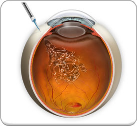
Retinal Photographs & Fluorescein Angiography
In order to most precisely diagnose Macular Degeneration, it may be necessary to have detailed color photographs of your macula and retina taken as well as a Fluorescein Angiogram (FA). After a fluorescent dye is injected into your arm, a series of photographs will be taken in rapid succession using a high-speed digital retinal camera as the dye passes throughout the retinal blood vessels. From these pictures it will be possible to see any fluid leakage or new blood vessel growth beneath the retina which will tell us whether certain types of treatments such as VEGF Inhibitor Injections might help stabilize your vision and prevent vision loss. Today, thanks to the advances in treating Wet Macular Degeneration, if caught early, it may be possible to avoid suffering significant vision loss.
Retinal Computer Imaging
We also use the advanced computer imaging technology called OCT or Optical Coherence Tomography that creates digital images of the retina and macula through the use of special beams of light in order to create a contour map of the retina and macula and detect microscopic changes in thickness or the accumulation of fluid beneath the retina or macula. This technique is similar to the CT Scans used to study organ systems and tissues throughout your body.
Vascular Endothelial Growth Factor (VEGF) Inhibitors
Lucentis® & Eylea® Injections
A specific protein called “Vascular Endothelial Growth Factor” (VEGF) causes the growth of new blood vessels or “neovascularization” to occur in the eye. From this work, drugs that can be injected into the eye in order to slow or stop the growth of new blood vessels have been developed. Two drugs, Eylea® and Lucentis® have been developed and FDA approved with specific indications to treat Wet Macular Degeneration. Each of these drugs works by inhibiting Vascular Endothelial Growth Factor (VEGF) so that there is little or no stimulus to grow new blood vessels in the Retina.
Eylea® and Lucentis® Injections are intravitreal injections-that means an injection that is placed directly into the Vitreous of the eye. Generally they need to be repeated every four to six weeks. Clinical studies of these anti-VEGF Injections indicate that when given to patients who have evidence of new blood vessel formation monthly, over 90% of patients will maintain their vision.
It is believed that nutrition may play a role in the likelihood of developing Macular Degeneration. Studies indicated that people who have a diet that is rich in fruits and vegetables-particularly green leafy vegetables-have a considerably lower incidence of Macular Degeneration. The Age Related Eye Disease Study (AREDS), which was sponsored by the National Eye Institute, showed that taking high levels of antioxidants and Zinc could reduce the risk of developing Age Related Macular Degeneration by about 25%. This is not a cure, but we need to consider this information as a possible way to help patients who are at risk for Age Related macular Degeneration prevent vision loss.
NOTE: A VERY SPECIFIC FORMULATION WAS USED IN THIS STUDY Before patients begin taking any course of vitamin or antioxidant supplements, you should fully discuss the risks and benefits with your Ophthalmologist, who in consultation with your family physician or Internist, will determine whether this is safe and effective for you to try.
